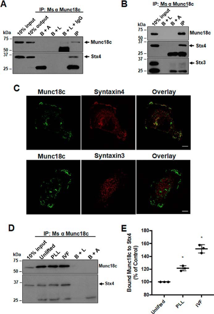
Figure 1.

Analysis of Stx4 and Munc18c association in MDA-MB-231 cells. A, Munc18c immunoprecipitates were probed for Munc18c (arrow) and Stx4 (arrow). B+A, beads plus antibody; B+L, beads plus lysate; B+L+IgG, beads plus lysate plus unrelated IgG; IP, immunoprecipitation; Ms, mouse. B, Munc18c immunoprecipitates were probed for Munc18c, Stx4 (arrow), and Stx3 (arrow). C, cells were grown for 4 h on glass coverslips, fixed, permeabilized, and stained for either Munc18c and Stx4 or Munc18c and Stx3. A representative confocal section from the ventral region of a cell is shown. Scale bars = 10 μm. D, analysis of Stx4 and Munc18c association during invadopodium formation (IVF). Cells were seeded onto coverslips coated with PLL or gelatin, incubated for 4 h, lysed, and analyzed by immunoprecipitation/Western blotting. Munc18c immunoprecipitates were probed for Munc18c and Stx4 (arrow). E, quantification of Stx4 co-immunoprecipitated with Munc18c. Percent of controls are from three independent experiments ± S.D. Asterisks denote values significantly different from control unlifted cells (*, p < 0.05). All data represent three or more biological replicates with at least three technical replicates.
