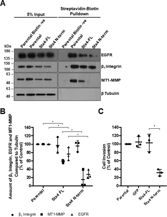
Figure 7.
Stx4 N-terminal peptide expression reduces cell surface levels of MT1-MMP and EGFR as well as cellular invasion. A, parental MDA-MB-231 cells and stable cell lines expressing GFP, GFP-Stx4-FL, or GFP–Stx4–N-term were plated onto gelatin for 4 h, exposed to biotin, and then lysed and analyzed by precipitation with streptavidin beads. Cell surface protein levels of β1 integrin, MT1-MMP, EGFR, and β-tubulin were assessed by Western blotting. Parental biotin −ve indicates cells exposed to buffer only. B, densitometric analysis of the amount of β1 integrin, EGFR, and MT1-MMP in streptavidin precipitations, as in A, relative to control. C, parental, GFP, GFP-Stx4-FL, and GFP–Stx4–N-term stable cells were serum-starved for 24 h, seeded onto transwell membranes coated with Matrigel or uncoated membranes (control), and allowed to invade for 20 h. Percentages of cells invading are shown from experiments in which at least 10 fields of view were counted per treatment. All data represent percent of control ± S.D. Asterisks denote values significantly different from control (*, p < 0.05). All data represent three or more biological replicates with at least three technical replicates.

