Figure 1.
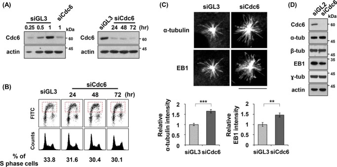
Cdc6 depletion increases microtubule regrowth from centrosomes. A, asynchronously grown U2OS cells were transfected with control GL3 or Cdc6-specific siRNA (see “Experimental procedures”) for 24 h or the indicated times, and then subjected to immunoblot analysis. The lysates of the control siRNA-treated cells were loaded with the indicated, relative volumes. Actin served as an internal control. B, FACS analysis was performed with BrdU (lower top) and propidium iodide (lower bottom) staining of the Cdc6-depleted cells for the indicated times after transfection. S phase cells are indicated in the dashed boxes. The proportions of replicating S phase cells are shown below the FACS profiles. siGL3, control siRNA GL3; siCdc6, Cdc6-specific siRNA. C, microtubule regrowth assays were performed with cells treated with the indicated siRNA for 24 h, as described under “Experimental procedures.” The cells, after incubation on ice for 1 h to depolymerize the microtubules, were incubated in a fresh medium at 37 °C for 15 s, and then fixed in the PEM + Fixative buffer. Microtubules were immunostained with antibodies specific to α-tubulin or EB1. Centrosomal intensities of α-tubulin and EB1 were densitometrically determined, and relative fluorescent intensities of α-tubulin and EB1 were plotted. Values represent mean ± S.D. of at least 100 cells in each of three independent experiments (**, p < 0.01; ***, p < 0.001). Scale bar, 10 μm. D, the indicated proteins were detected in immunoblots with antibodies specific to each protein.

