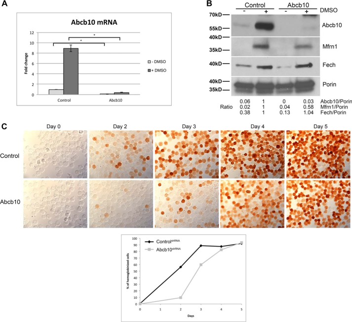
Figure 2.
Abcb10 shRNA-stable MEL cells show decreased hemoglobinization. A, MEL cells stably expressing control shRNA or Abcb10-specific shRNA were treated with 1.5% DMSO for 3 days, and mRNA was isolated. qRT-PCR for Abcb10 and actin were performed. Error bars, S.E. B, mitochondria were isolated from cells as in A and lysed, and Western blot analysis was performed using rabbit anti-Abcb10, rabbit anti-Mfrn1, mouse anti-Fech, and mouse anti-Porin followed by HRP-conjugated goat anti-rabbit or anti-mouse IgG. Blots were quantified using NIH ImageJ with porin as a loading control and the ratios of Abcb10, Mfrn1, and Fech to Porin were determined with control shRNA MEL cells normalized to 1. An example blot with its quantification is shown. C, MEL cells stably expressing control shRNA or Abcb10-specific shRNA were treated with 1.5% DMSO for 0–5 days and stained for hemoglobin using o-dianisidine. Quantification of hemoglobinization of a representative experiment is shown (n = 100–200 cells/time). (The presence of a pink to red signal was considered positive in the quantification). *, p ≤ 0.05.

