
Figure 4.
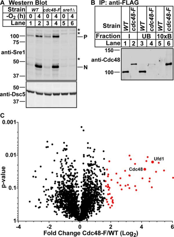
Identification of Cdc48-binding proteins in S. pombe. A, Western blottings, probed with monoclonal anti-Sre1 IgG (5B4) and polyclonal anti-Dsc5 IgG (for loading), of lysates treated with alkaline phosphatase for 1 h from wild-type cells, cdc48-5xFLAG (cdc48-F) cells, or sre1Δ cells grown for 0 or 4 h in the absence of oxygen. P and N denote precursor and cleaved N-terminal transcription factor forms, respectively. Asterisks denote non-specific bands. B, Cdc48 was immunoprecipitated (IP) from wild-type or cdc48-5xFLAG cells using monoclonal anti-FLAG M2 IgG as described under “Experimental procedures.” Input, unbound, and 10-fold enriched bound fractions were analyzed by Western blotting using polyclonal anti-Cdc48 IgG. The blot is representative of four biological replicates. C, volcano plot of all proteins identified during TMT mass spectrometry of Cdc48–5xFLAG bound proteins. Points in red are proteins with enrichment >2 S.D. from the mean in the Cdc48–5xFLAG versus wild-type samples. p values were calculated using two-way ANOVA with quantile normalization for three biological replicates.