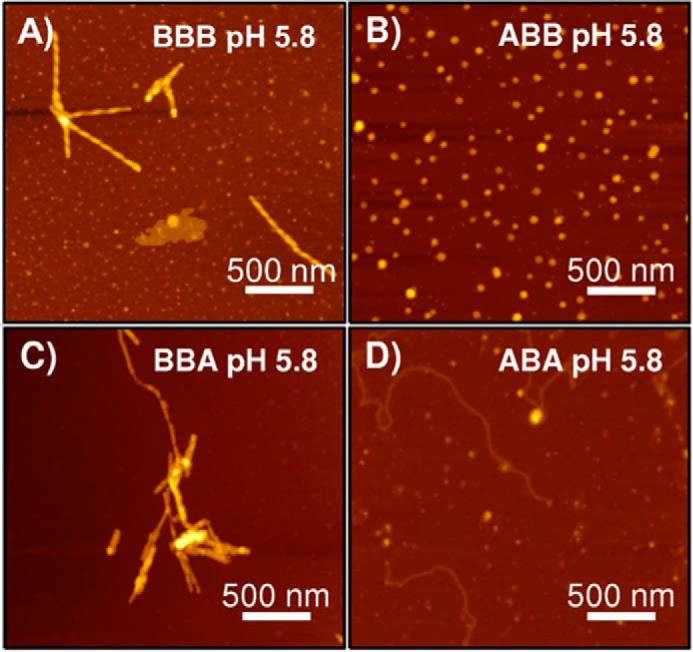
Figure 5.

AFM images of low pH 5.8 ABX and BBX fibrils. Non-contact mode AFM images for low pH samples of chimeric constructs with a βS NAC domain in a 2.5 × 2.5 μm spot size are shown. A, WT βS; B, ABB; C, BBA; and D, ABA chimeric proteins. Scale bar, 500 nm.
