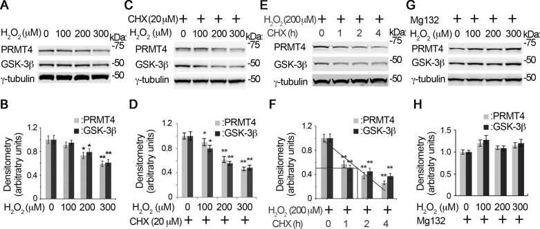
Fig. 2.
H2O2-induced PRMT4 instability is via proteasomal degradation. A–F: time and concentration studies of PRMT4 degradation under H2O2. MLE12 cells were treated with H2O2 with a range of concentrations in the absence (A and B) or presence of cycloheximide (CHX) for 4 h (C and D), or cells were treated with CHX for various time durations in the presence of 200 μM H2O2 (E and F). The cell lysates were subjected to PRMT4, GSK-3β, and γ-tubulin immunoblotting analysis. The densitometry results of A, C, and E were plotted, and the half-life of the proteins were calculated (B, D, and F). G: MLE12 cells were treated with diverse concentrations of H2O2 (0, 100, 200, and 300 μM) in the presence of MG132 (20 μM) for 4 h. Cell lysates were subjected to PRMT4, GSK-3β, and γ-tubulin immunoblotting analysis. H: the densitometry results of G were plotted. Results are representative of 3 independent experiments *P < 0.05 or **P ≤ 0.01 vs. corresponding control.

