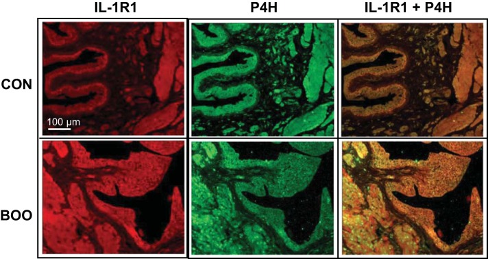
Fig. 6.
Coimmunofluorescence staining of bladder sections for IL-1R1 and P4H. Tissue sections from bladders were stained with antibodies to IL-1R1 and P4H (both at 1:50 dilution) as described. Secondary antibodies were conjugated to Texas red and Alexa Flour 488 as described. Staining was visualized, and at right is an overlay of the two stains where yellow indicates colocalization. It should be noted that this is the same section used in Fig. 4, left, and the P4H stain is the exact image.

