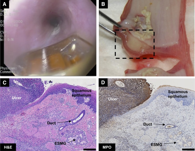
Fig. 3.
Assessment of radiofrequency ablation (RFA)-generated wound in porcine esophagus. A: endoscopic view of uninjured porcine esophagus with RFA probe. B: gross appearance in cross section of esophageal injury 48 h postablation with extensive ulceration and mild edema. C: hematoxylin and eosin (H&E) staining 48-h postablation, with a demarcation between ulcerated (left) and healthy tissue (right) where an ESMG and duct are present. D: myeloperoxidase (MPO) staining for neutrophils demonstrated evidence of ulceration on the top left of the image and a clear demarcation at the edge of the injured area. Scale bar = 200 µm.

