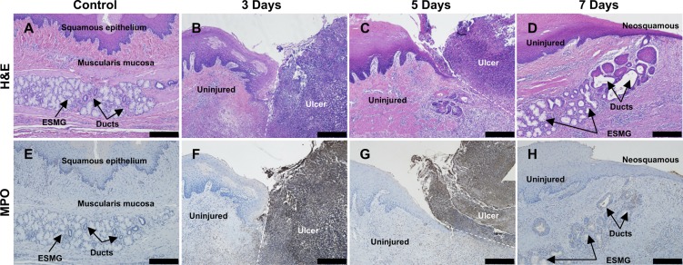
Fig. 4.
Areas of RFA injured esophageal tissue at 3, 5, and 7 days were compared with control using both H&E and MPO. A: control H&E with normal appearance of an ESMG with overlying squamous epithelium. B: at 3 days, a large ulcer was present (right) with clear demarcation between the uninjured tissue (left) and ablated, ulcerated tissue (right). C: at 5 days, a small area of neosquamous epithelium (left) appeared underneath ulcerated tissue (right) above an ESMG at the junction of injured and uninjured tissue. D: 7 days postablation, neosquamous epithelium appeared along with an ESMG with the ductular phenotype and areas of squamous islands near the neosquamous epithelium. E: uninjured control tissue did not demonstrate MPO-positive cells. F: 3 days after injury, dark MPO staining was present on the right in the ablated area, with little to no staining present in the uninjured tissue (left). G: at 5 days, MPO-stained tissue (right) separated from uninjured, repairing epithelium (left). H: neosquamous epithelium and associated ESMG with little to no positive MPO cells, indicating a reduction in inflammation and resolution of the ulcer. Scale bar = 200 µm.

