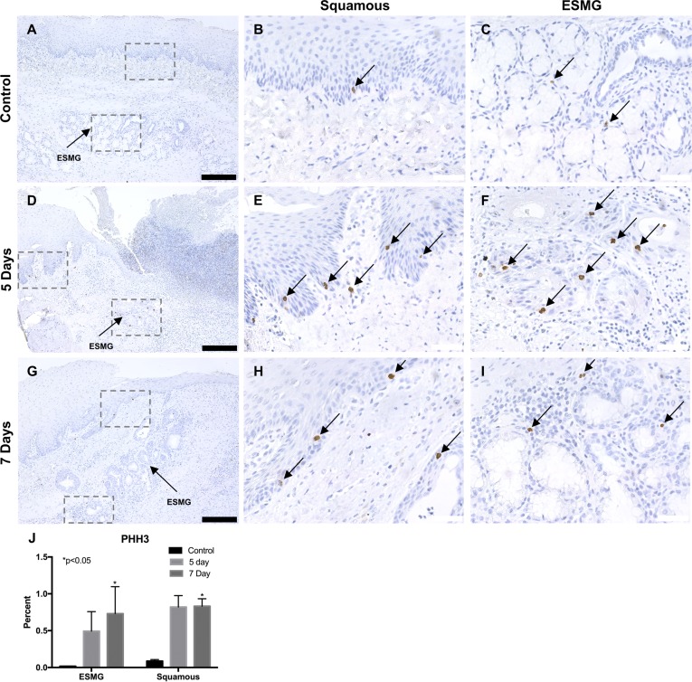
Fig. 5.
Phosphohistone H3 (PHH3), an M-phase marker for proliferation, was used to detect proliferating cells in the porcine esophagus. A–C: in uninjured esophagus, cells were rarely positive for PHH3 in the squamous epithelium (B) and ESMGs (C). D-F: 5 days postablation (n = 3), a marked increase of PHH3-positive cells was noted in both the squamous epithelium (E) and ESMGs (F). G–I: 7 days postablation (n = 6), PHH3-positive cells were present in the squamous epithelium (H) and ESMGs (I). J: after quantification, compared with uninjured control, PHH3 increased significantly in ESMGs 7 days postablation (*P < 0.05) and increased significantly in squamous epithelium 7 days postablation (P < 0.05). Scale bar = 200 µm (black) and 40 µm (white). Percentages reported as means ± SE.

