Fig. 6.
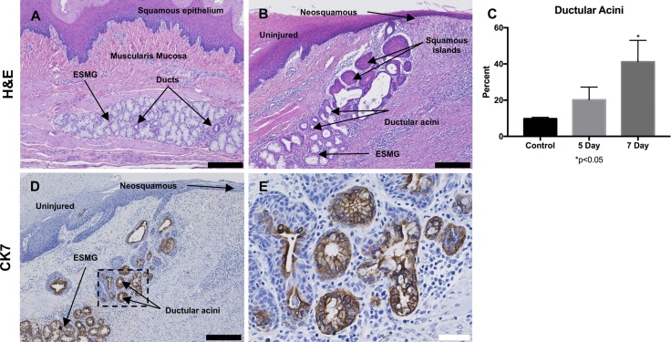
A ductular phenotype was present in injured porcine ESMGs. A: control ESMGs contained mucinous acini with a few small normal ducts to collect acinar secretions. B: 7 days postablation, the ESMGs exhibited an increased ductular phenotype rather than mucinous acini within the ESMGs, with additional squamous islands near the lumen. C: when acinar phenotype was counted (ductal vs. mucinous) and compared with uninjured control, 5 days postablation the ductular phenotype nearly doubled compared with control, and it quadrupled at 7 days to 41.16 ± 11.85% compared with control (*P < 0.05). D: CK7, a ductal marker, stained strongly positive in 7 days postablation ESMGs. E: CK7 in higher magnification image demonstrating acini with mixed phenotype, dilation of the acini, loss of mucin. Multiple layers are present in some ducts, particularly near the overlying repairing squamous epithelium. Scale bar = 200 µm (black) and 40 µm (white). Percentages reported as mean ± SE.

