Fig. 7.
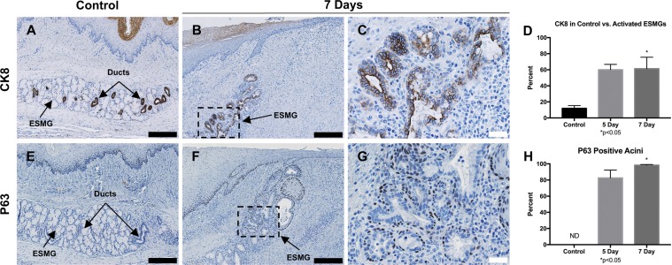
Epithelial markers P63 and CK8 in RFA-injured tissue. A: CK8 expression in control ESMGs had strong expression within the ducts. B and C: after injury, expression of CK8 was strongly present in the ductular acini with a notable expansion. D: in control tissue, 11.99 ± 3.39% of the control ESMGs consisted of CK8-positive ducts. Compared with uninjured controls, CK8 expression increased to 60.13% ± 6.54% in ESMGs 5 days postablation, and to 61.41 ± 14.23% (*P < 0.05) in ESMGs 7 days postablation. E: P63 expression was present on the basal cells in the ducts contained within ESMGs but not detected (ND) in the acini of the uninjured ESMGs. F and G: P63 expression remained active in the ducts associated with ESMGs postablation, and P63-positive cells were found in the acini of injured ESMGs. H: compared with uninjured controls, P63-positive cells were present in 82.64 ± 9.65% of acini in injured ESMGs 5 days postablation, and significantly increased to 98.33 ± 0.88% (P < 0.05) in injured ESMGs 7 days postablation. Scale bar = 200 µm (black) and 40 µm (white). Percentages reported as mean ± SE.

