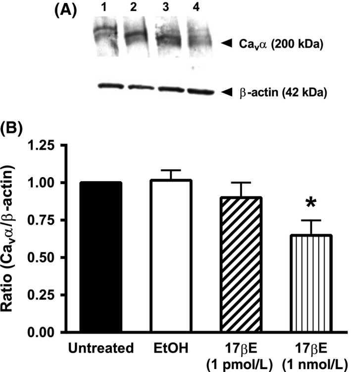
Figure 4.

Exposure to 17βΕ for 24 h decreases the expression of the α 1C pore‐forming subunit (Cav α) of the Cav1.2 channel in coronary arterial strips. (A) Diffuse immunoreactive band corresponding to the Cav α protein; the short (~200 kD) and long (~240 kD) forms of Cav α are revealed as a doublet in some blots. β‐Actin (42 kDa) was used as an internal standard. Protein lysates were obtained from cultured arteries exposed for 24 h to the following conditions: lane 1 = untreated; lane 2 = EtOH (0.1%); lane 3 = 1 pmol/L 17βΕ; lane 4 = 1 nmol/L 17βΕ. B, The densitometric ratio corresponding to Cav α in arterial lysates exposed for 24 h to EtOH or 1 nmol/L 17βΕ compared to untreated arteries. Results indicate that 1 nmol/L 17βΕ for 24 h decreases Cav α protein abundance. Data represent mean ± S.E. (n = 5 pigs) and analyzed using one‐way ANOVA followed by Bonferroni's post hoc analysis. There was no difference in β‐actin expression between groups.
