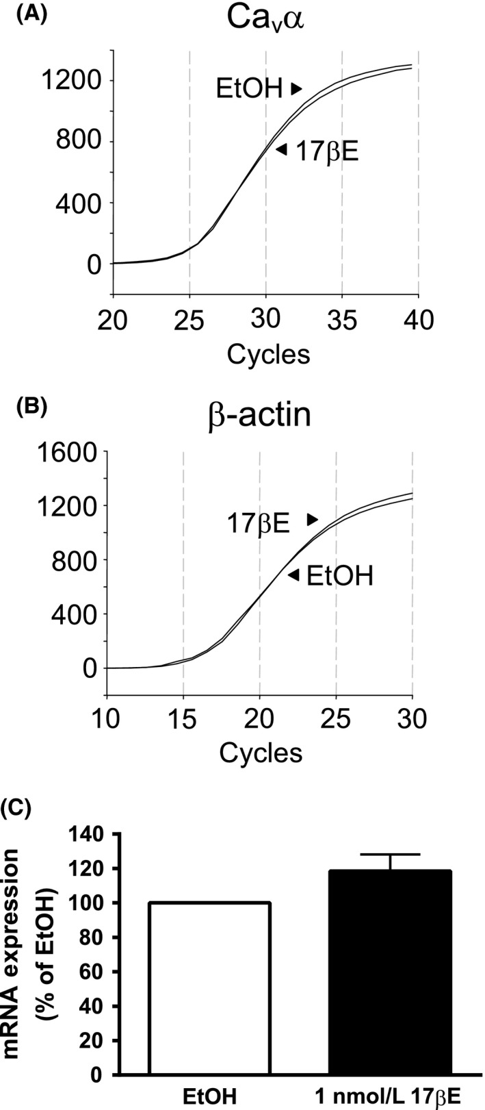
Figure 5.

Exposure of arterial segments to 1 nmol/L 17βE for 24 h failed to alter expression of Cav α transcript. (A and B) Real‐time PCR amplifications of Cav α (A) and internal standard β‐actin (B) are similar between arteries cultured for 24 h in RPMI media containing 1 nmol/L 17βΕ or its solvent (EtOH). Relative fluorescence units. (B) Abundance of Cav α transcript is not significantly different between 17βΕ and EtOH‐treated arteries as analyzed by the ΔΔCt method. Data are expressed as percent (%) transcript expression in EtOH (n = 6 pigs).
