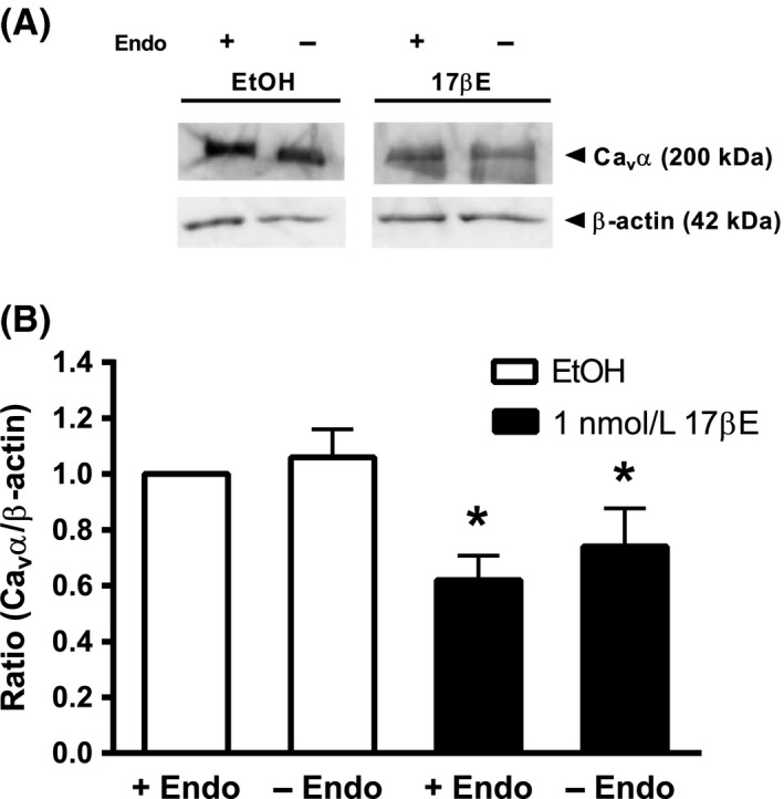
Figure 7.

17βΕ decreases expression of the Cav α pore‐forming subunit independently of endothelium. Upper panel shows a representative anti‐Cav α immunoblot from arteries with intact (+Endo) or denuded (‐Endo) endothelium, which were cultured for 24 h in RPMI media containing 1 nmol/L 17βΕ or ethanol (EtOH). 17βΕ decreased the densitometric ratio in intact and denuded arteries. β‐actin served as an internal standard. The images are from different parts of the same gel. The Cav α and β‐actin groupings were imaged separately. Band intensity between groups was normalized to β‐actin and expressed as a densitometric ratio of the EtOH+Endo group. Data represent mean ± SE (n = 7 pigs). *P < 0.05, EtOH ± Endo versus 17βΕ ± Endo using a two‐way ANOVA (without replication) followed by Tukey HSD post hoc analysis.
