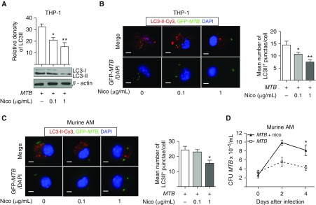
Figure 3.
Nicotine inhibits autophagosome formation in MTB-infected macrophages. (A) THP-1 cells were infected with MTB alone or with 0.1 and 1 μg/ml of nicotine (Nico) for 18 hours, whole-cell lysates prepared, proteins separated by SDS-PAGE, and immunoblotted for microtubule-associated protein light chain-3-I (LC3-I) and LC3-II. (B) Representative immunocytofluorescence of LC3-II–Cy3+ autophagosomes in THP-1 cells infected with GFP-MTB alone or with 0.1 and 1 μg/ml nicotine. To quantify the number of autophagosomes per cell, the numbers of LC3-II–Cy3+ punctae were counted from at least 100 random cells/well in duplicate wells for each condition. Data shown are the mean (±SEM) of three independent experiments. (C) Representative immunocytofluorescence of LC3-II–Cy3+ autophagosomes in murine alveolar macrophages (AMs) infected with GFP-MTB alone or with 0.1 and 1 μg/ml nicotine. Quantitation of the number of autophagosomes per cell is as described in B. Data shown are the mean (±SEM) of three independent experiments. (D) Murine AMs were infected with MTB alone or with 1 μg/ml nicotine. At 1 hour and 2 and 4 days after infection, macrophage-associated MTB was quantified. Data shown are the mean (±SEM) of two independent experiments performed in duplicates. Scale bars: 5 μm. *P < 0.05, **P < 0.01.

