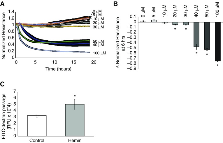
Figure 1.
Hemin decreases the transendothelial electrical resistance (TER) of a human lung microvascular endothelial cell (hLMVEC) monolayer in a concentration-dependent manner and increases fluorescein isothiocyanate (FITC)-dextran passage. hLMVECs were grown to confluency on gold electrodes. Resistance to an electrical current applied to the electrode was measured and used as a marker for the degree of barrier-enhancing cell–cell attachment after treatment with varying concentrations of hemin (100 μM, 50 μM, 40 μM, 30 μM, 20 μM, 10 μM, and 5 μM) versus vehicle-treated controls. The plot in A depicts the average electrical resistance normalized to baseline resistance over time after hemin treatment in n = 3 experiments. Hemin was added at time 0. Error bars are ± SD. The bar graph in B shows the maximal percent decrease in resistance after 6 h of hemin exposure versus hemin concentration/vehicle-treated controls. In separate experiments, hLMVECs were grown to confluency on Transwell inserts with 3 μm pores in media that contained 3 kD FITC-dextran. After treatment with either vehicle control or 40 μM hemin, media from below the monolayer was sampled and analyzed by fluorometry for the presence of FITC-dextran. The bar graph in C depicts the relative fluorescence units (RFU) of media taken from below control versus hemin-treated monolayers in n = 3 experiments. Error bars are ± SD. *Indicates significant difference from control (Student’s t test, P < 0.05).

