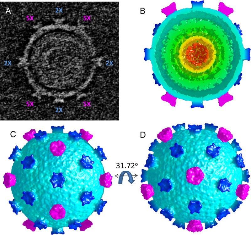
FIG 7.
Cryo-electron tomogram of a single, icosahedrally averaged MTIV particle. (A) A single slice through the center of the unaveraged particle rotated into a standard icosahedral orientation (2-fold axes along x, y, and z). The locations of the 2-fold (2×) (magenta) and 5-fold (5×) (blue) axes lying in the central plane are indicated. (B) A single hemisphere of the icosahedrally averaged particle (in a standard orientation; comparable to panel A) reveals the outer surface layer (cyan) surrounding an inner shell (green) that, in turn, encases three additional layers of concentric density (green, yellow, orange) indicative of packaged DNA. (C) A surface view down the icosahedral 2-fold axis (standard orientation). The turrets at the icosahedral 5-fold axes (magenta) and the approximately 6-fold symmetric turrets on the 2-fold axes (dark blue) are shown. (D) The particle is rotated 31.72° about the horizontal axis to show the view down an icosahedral 5-fold axis.

