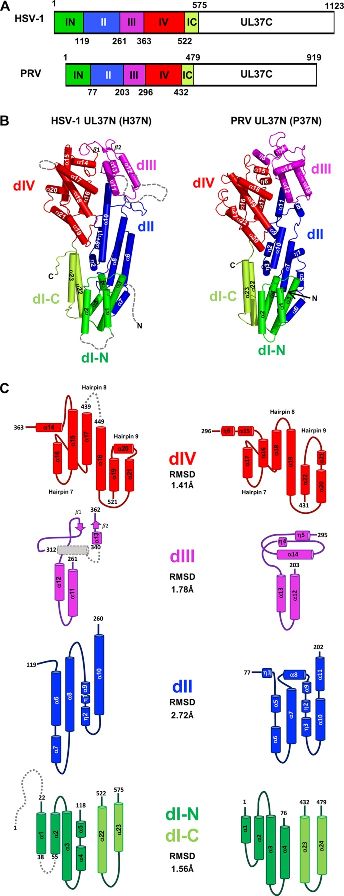
FIG 1.
Domain organization in UL37N structures from HSV-1 and PRV. (A) Linear diagram of UL37 from HSV-1 and PRV with domains colored and domain boundaries labeled. (B) Crystal structure of UL37N from HSV-1 (H37N, left) and PRV (P37N, right) colored by domain as in panel A. Secondary structure elements are numbered sequentially. The orientation was chosen to show all secondary structure elements. (C) Topology diagrams of individual domains. Helices are shown as cylinders and beta strands as arrows. The color scheme is the same as in panels A and B.

