FIG 6.

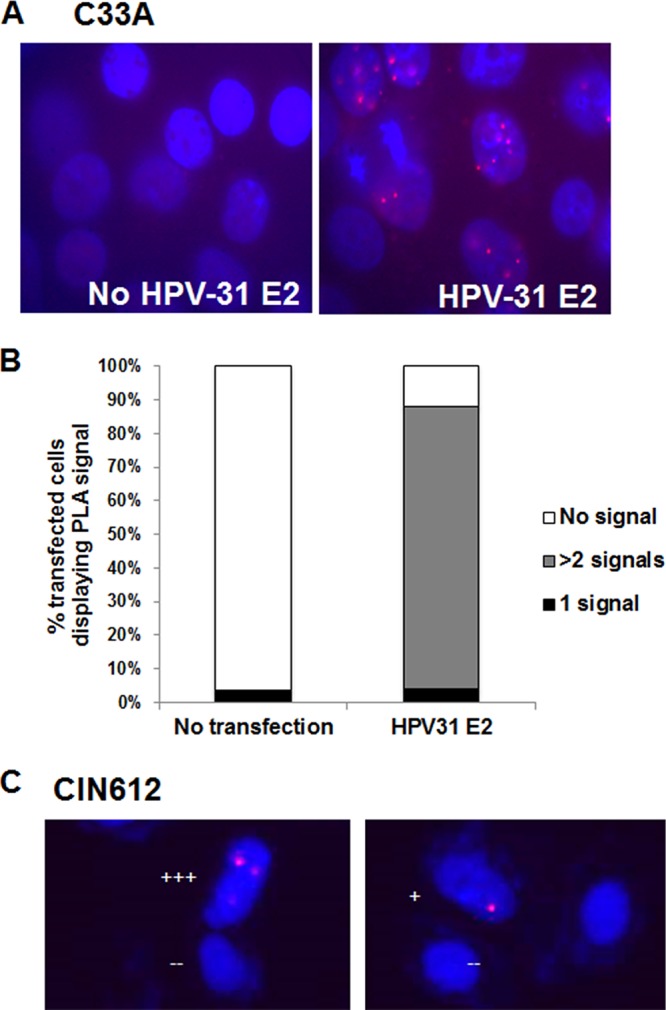
FGFR3 interacts with E2 within the nucleus. (A) C33A cells were transfected with and without FLAG-HPV-31 E2. PLA was completed with M2 (mouse) and FGFR3 (rabbit) antibodies. Red foci are an indication that the FLAG and FGFR3 proteins are within close proximity of each other. Red, focus formation; blue, DAPI. (B) FGFR3 and FLAG-HPV-31 E2 PLA foci were counted without and with FLAG-HPV-31 E2 expression. The percentages of cells displaying 0, 1, or more than 2 signals are shown. (C) CIN612-9E cells were transfected with and without FLAG-HPV-31 E2. PLA was completed with M2 (mouse) and FGFR3 (rabbit) antibodies. Red foci are an indication that the FLAG and FGFR3 proteins are within close proximity of each other. Red, focus formation; blue, DAPI. Each plus sign indicates the number of foci.
