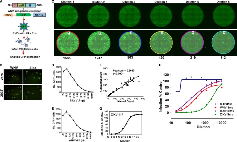
FIG 2.
RVP-based microneutralization assay for ZIKV, using a 96-well plate format and a GFP readout. (A) Strategy for generation of ZIKV or WNV reporter virus particles. 293T cells were cotransfected with ZIKV/WNV C-prM-E along with the WNV subgenomic replicon construct Rep/GFP. Culture supernatants were harvested at 48 h posttransfection and used to infect 293T/Vero cells, and GFP expression was analyzed as a measure of virus infection. (B) 293T and Vero cells were infected with ZIKV or WNV RVPs, and infection was analyzed by fluorescence microscopy to detect GFP-positive cells. (C) Vero cells were infected with serial dilutions of ZIKV reporter virus-like particles in 96-well plates. Cells were fixed at 72 h postinfection, and images of whole wells were captured via fluorescence microscopy. Representative fluorescence images of whole wells infected with 6 serial dilutions (dilutions 1 to 6) of ZIKV RVPs are depicted. The top panel shows raw images acquired by fluorescence microscopy. The bottom panel shows the same images analyzed using the automated NIS Elements software, which marks GFP-positive cells by using the cell count function. The number below each well represents the number of GFP-positive cells in that well. Vero cells were infected with serial dilutions of ZIKV (D) or WNV (E) reporter virus particles in 96-well plates. The experiment was conducted with 6 technical wells infected for each dilution. The number of GFP-positive cells was determined at 72 h postinfection as described for panel C. The data show small variations between the 6 wells for each RVP dilution. (F) Quantitation of GFP-positive cells infected with ZIKV RVPs by using the automated software from Nikon versus manual counting. Representative data show a high degree of correlation between the two methods. ZIKV-117 antibody (G) or the indicated antibodies/sera (H) were serially diluted in DMEM and incubated with a predetermined amount of ZIKV RVPs for 1 h at room temperature. Subsequently, the virus-serum mixtures were added to Vero cells. The cells were incubated for 72 h, after which images were acquired and the number of GFP-positive cells quantitated as described above. The assay was conducted in technical triplicates for the ZIKV-117 antibody and ZIKV sera and in duplicates for others. Data representative of 1 of 3 independent experiment are shown.

