FIG 1.
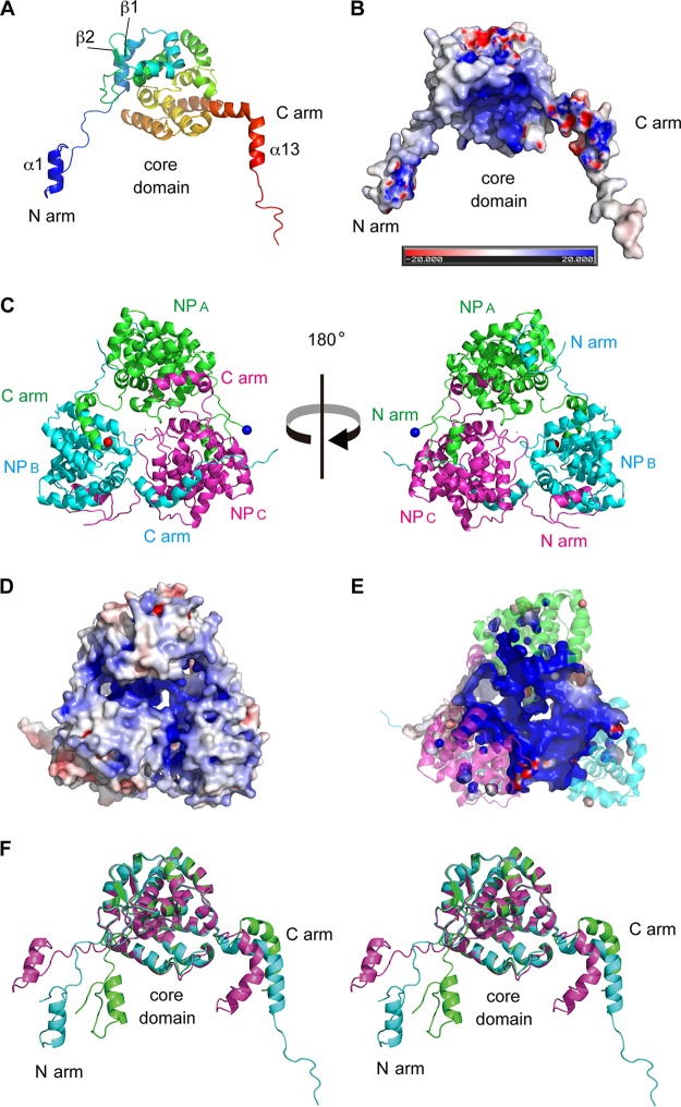
Crystal structure of the TSWV N protein. (A) Structure of the TSWV N protein protomer (NPB). The N-terminal arm (N arm), central core domain (core domain), and C-terminal arm (C arm) are indicated. (B) Analysis of the electrostatic surface of TSWV NPB viewed from the same direction as in panel A. Blue and red represent positive and negative potentials, respectively. The color scale ranges between −20 kBT and +20 kBT, where kB is Boltzmann's constant and T is temperature. (C) Cartoon representation of the TSWV N protein trimeric ring. NPA, NPB, and NPC are depicted in green, cyan, and magenta, respectively. The N and C termini of NPA are marked with blue and red spheres, respectively. (D) Analysis of the electrostatic surface of the TSWV N protein trimer viewed from the same direction as in panel C (right). (E) Transverse section of the structure in panel D at the level of the positively charged cleft. (F) Stereo view of the superposition of NPA, NPB, and NPC. Each N protein molecule is shown in the same color as that described above for panel C.

