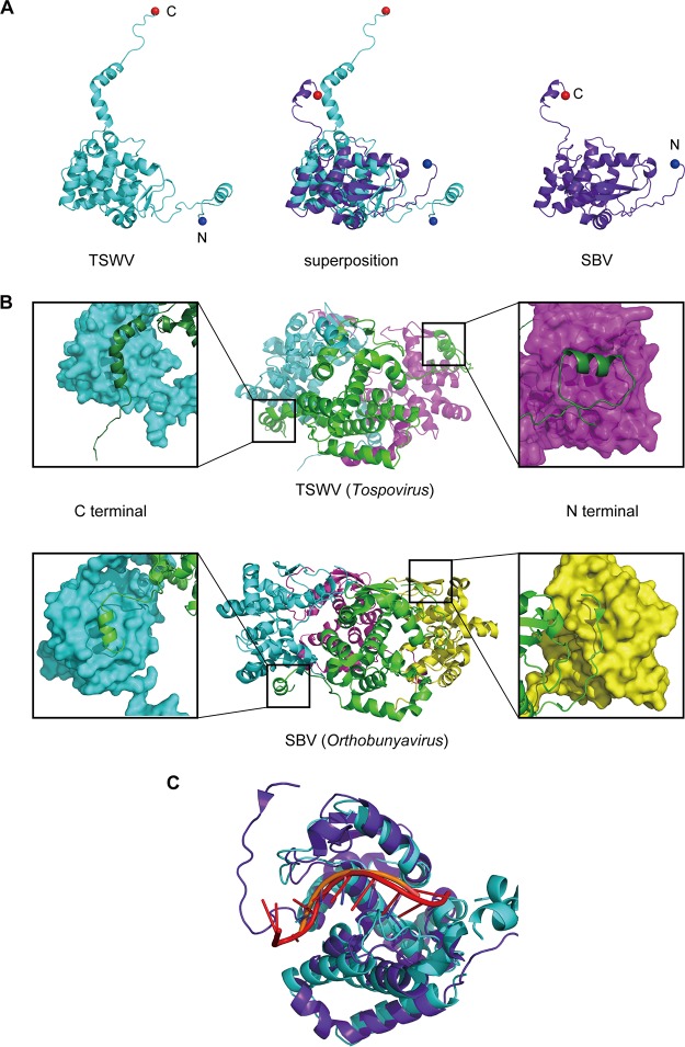
FIG 4.
Comparison of N protein structures of TSWV (Tospovirus) and SBV (Orthobunyavirus). (A) Protomer structures of the TSWV N protein (left) and the SBV N protein (PDB accession no. 4JNG) (right) and the superposition of both structures (middle). (B) Magnified views of the arm-to-core interaction within the TSWV (top) and SBV (bottom) N proteins are shown for each side. In the magnified views, NPA is shown as a green cartoon, whereas the adjacent N protein molecule is depicted as a molecular surface. (C) Superposition of the TSWV N-RNA complex (cyan, TSWV N; orange, RNA) and the SBV N-RNA complex (purple, SBV N; red, RNA).

