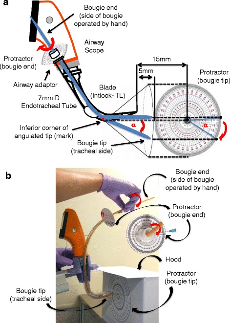
Fig. 1.

Experimental protocol for the bench study. According to the instruction manual [3], a 7.0 mm internal diameter standard endotracheal tube is inserted into the guiding channel of an Airway Scope blade, the tip of which is set 15 mm from the circular protractor that stands vertically on a table (a). The anesthesiologist marks the inferior corner of the angulated tip of the bougie then, with the angulated tip directed towards 12 o’clock, inserts the bougie into the tube, locates the tip 5 mm beyond the blade tip, and aligns the mark with the center of the protractor on the monitor screen of the AWS (a). The power switch of the Airway Scope is turned off. The anesthesiologist then rotates the bougie end (side of bougie operated by hand) clockwise or counterclockwise to an angle of 0°–180° in 45° increments while looking at the protractor that is set on the airway adaptor of the endotracheal tube (b). The bougie tip (tracheal side) is not visible because of the hood (b). The angle of the bougie tip created by rotating its end is measured. α: rotation angle of the bougie tip
