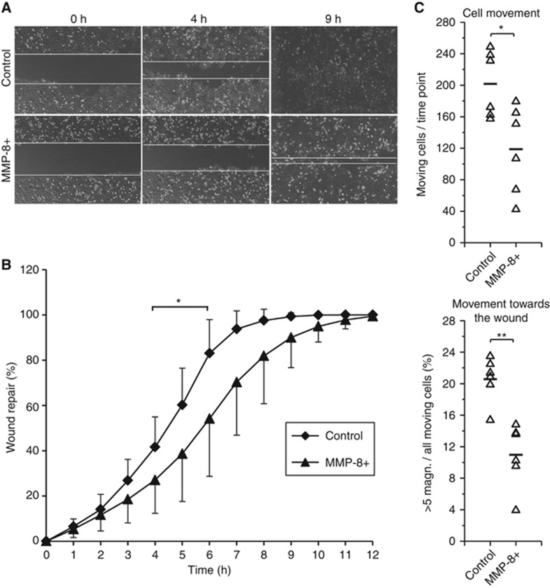
Figure 2.
MMP-8 reduced the migration of HSC-3 cells. After removing the ibidi inserts, the migrating cells were photographed for 12 h (10 min intervals) (A). The open areas were measured using Fiji software and the results were calculated as a percentage of area closure (mean and standard deviation are shown) (B). Phase contrast time-lapse images were acquired at multiple stage positions using a motorised stage with Cell^P software and the results were analysed using a Kalman filter-based tracking approach with MATLAB (C). *P<0.05, **P<0.01. Six replicates were analysed for each cell group in all experiments.

