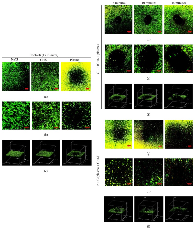
Figure 7.
Confocal laser scanning microscope (CLSM) images of biofilms at titanium coupons in controls and combination treatment group: C + P and P + C. The titanium coupons (both treated and control) containing biofilms were subjected to CLSM and representative images were taken from the coupon. (b), (e), and (h) show 2D confocal images of control and treated (C + P and P + C) biofilms and (c), (f), and (i) represent the 3D volume image of the control and treated (C + P and P + C) biofilms, respectively. (a), (d), and (g) are merged images with multiple images. (b), (e), and (h) are a single image at the treated region. Green coloration and yellow coloration represent live and dead cells as stained by SYTO9 and PI (propidium iodide), respectively. The round black region at the center is the area directly treated by the plasma jet. The scale bar is 250 µm.

