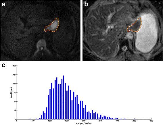
Fig. 2.

A 74-year-old woman with gastric carcinoma pathologically staged as T3N1cM0. a Axial diffusion weighted image (b = 1000 s/mm2) showed the lesion with high signal intensity in the lesser curvature of stomach; (b) The outline of the lesion was automatically copied to the same location of the apparent diffusion coefficient (ADC) map at the same level as (a); (c) The histogram of ADC map, with a bin size of 50 × 10−6 mm2/s: ADCmean* = 1520.76, ADCmin* = 437, ADCmax* = 3502, ADC5%* = 957, ADC10%* = 1025, ADC25%* = 1194, ADC50%* = 1443, ADC75%* = 1777, ADC90%* = 2110 (note: * The unit for ADC value is ×10−6 mm2/s)
