
Fig. 6.
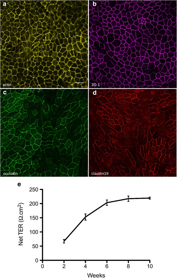
Tight junction proteins and the transepithelial resistance of iPSC-RPE. RPE cells were cultured on laminin-coated Transwell inserts. a Phalloidin labeling demonstrates the cortical arrangement of actin filaments in iPSC-RPE. b–d Immunostaining of the iPSC-RPE cells revealed surface expression of the tight junction proteins ZO-1 (b), occludin (c), and claudin19 (d). e To assess the barrier function of the iPSC-RPE cells, the resistance across monolayers of cells cultured on laminin-coated Transwell inserts was measured at 2-week intervals, following initiation of the cultures. The net TER was determined by subtracting the resistance across a laminin-coated Transwell insert lacking cells, and multiplying by the surface area of the insert (0.33 cm2). The net TER of the iPSC-RPE cells steadily increased between weeks 2 and 8, until it reached a maximal value of just above 200 Ω.cm2. Resistance recordings were made from four individual cultures. e Error bars represent the mean ± the SEM. Scale bars: a–d = 20 μm. ZO-1 zonula occludens-1, TER transepithelial resistance