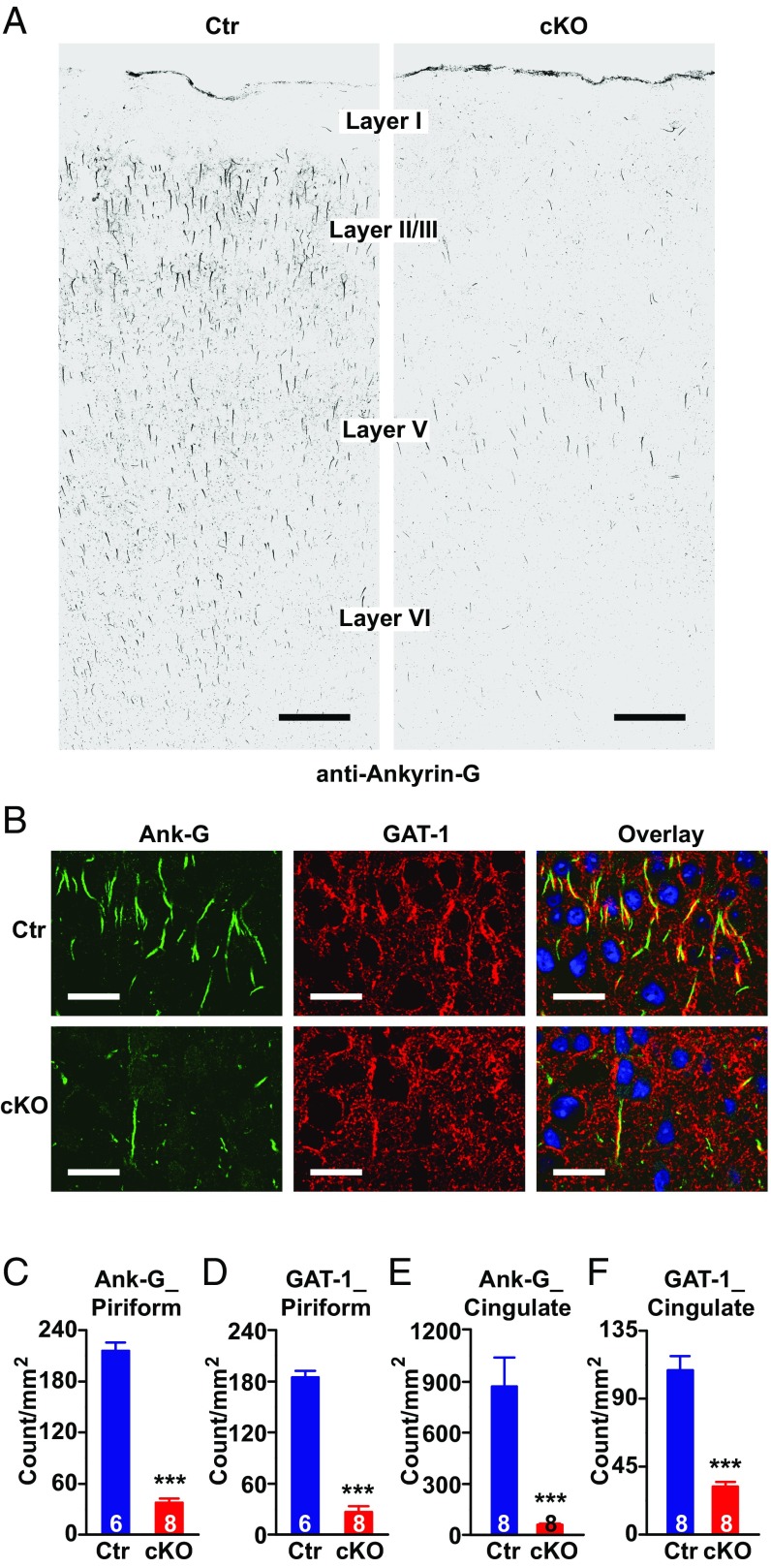
Fig. 1.
Genetic deletion of ankyrin-G in AISs of pyramidal neurons of mouse forebrain, and alteration of presynaptic GABA markers. (A) Confocal images of mouse motor cortex immunolabeled with anti–ankyrin-G antibody showing loss of ankyrin-G at AIS in Ank-G cKO mouse. (Scale bar: 100 µm.) (B) Confocal images of mouse piriform cortex labeled with anti–ankyrin-G (green) and anti–GAT-1 (red) antibodies in wild-type (Top) and Ank-G cKO (Bottom) mice. (Scale bars: 30 µm.) The nuclei are labeled with DAPI (blue). (C–F) Quantification of ankyrin-G (C and E) and GAT-1 (D and F) at AIS in mouse piriform cortex (C and D) and cingulate cortex (E and F). Bar graphs represent mean ± SEM; ***P < 0.001, Student t test. The number of animals per group is indicated at the bottom of each bar graph.

