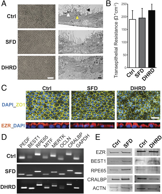
Fig. 1.
No difference in baseline RPE characteristics were seen in Ctrl, SFD, and DHRD hiPSC-RPE at D90 in culture. (A) Light and electron microscopy images of D90 SFD, DHRD, and Ctrl hiPSC-RPE cultures grown on Transwell inserts (Corning) showed the characteristic cobblestone RPE morphology, apical microvilli (black arrowhead), melanosomes (white arrowhead), and mitochondria (yellow arrowhead). (Scale bars: 50 μm, Left; 1 μm, Right.) (B) Transepithelial resistance measurement of D90 SFD, DHRD, and Ctrl hiPSC-RPE cultures was comparable to the proposed in vivo threshold, 150 Ω·cm−2 (31). Data are expressed as mean + SEM. (C) Immunocytochemical analyses demonstrated RPE-specific morphology, i.e., tight junction formation (ZO-1) and proper polarization (EZR: Apical), of cultured SFD, DHRD, and Ctrl hiPSC-RPE at D90. (Scale bar: 50 μm.) (D and E) RT-PCR (D) and Western blot (E) analyses showed robust expression of RPE characteristic genes and proteins in D90 hiPSC-RPE derived from SFD, DHRD, and Ctrl hiPSCs. GAPDH and ACTN served as loading controls in RT-PCR and Western blotting analysis, respectively. Note: The color palette in confocal images throughout the article has been altered to accommodate colorblind readers.

