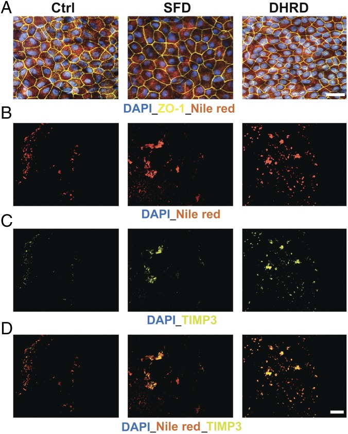
Fig. 3.
Transwell membranes underneath aged (D90) SFD and DHRD hiPSC-RPE show deposition of Nile red-stained TIMP3 containing lipid–protein complexes. (A) Nile red staining demonstrated uniform intracellular expression of neutral lipids in D90 Ctrl, DHRD, and SFD hiPSC-RPE. (Scale bar: 50 μm.) (B–D) Immunostaining of Transwell membranes after removal of the D90 hiPSC-RPE monolayer showed increased sub-RPE deposition of neutral lipids (B), TIMP3 (C), and colocalized neutral lipid-TIMP3 complexes (D) on the surface of Transwell membranes underlying patient SFD and DHRD hiPSC-RPE cultures compared with Ctrl hiPSC-RPE cultures. (Scale bar: 50 μm.) Of note, confocal images from the same experiment showing the same Transwell membrane are shown in B–D to emphasize the almost complete colocalization of TIMP3 and Nile red staining in sub-RPE deposits on Transwell membranes underlying SFD and DHRD hiPSC-RPE cultures.

