Fig. 5.
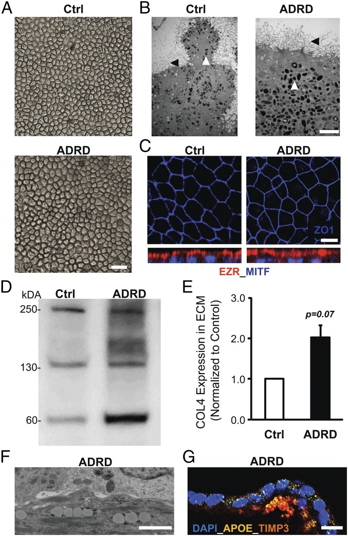
Increased accumulation of COL4 in ECM and the presence of TIMP3-APOE–positive deposits in aged (D90) ADRD hiPSC-RPE cultures. (A and B) Light microscopy (A) and electron microscopy (B) analysis at D90 showed similar cobblestone morphology in Ctrl vs. ADRD hiPSC-RPE cultures. Apical microvilli (black arrowhead), and melanosomes (white arrowhead) are seen in Ctrl vs. ADRD hiPSC-RPE cultures. (Scale bars: 100 µm in A; 5 µm in B.) (C) Immunocytochemical analyses demonstrated similar localization of the tight junction protein ZO-1 and the apical RPE cell marker EZR in Ctrl and ADRD hiPSC-RPE at D90. (Scale bar: 10 μm.) (D and E) Quantitative Western blot analyses demonstrated higher levels of COL4 protein in the ECM underlying Ctrl vs. ADRD hiPSC-RPE cultures at D90. Of note, data are presented as mean + SEM in the bar graph. Furthermore, COL4 bands at ∼250, 150, and 70 kDa are consistent with multi- and monomeric α1, α2 subunits and fragments of the human COL4 protein. (F and G) Electron microscopy (F) and immunocytochemical (G) analyses revealed lipid droplet accumulation and TIMP3-APOE–positive sub-RPE deposits in D90 ADRD hiPSC-RPE cultures. (Scale bars: 1 µm in F and 50 µm in G.)

