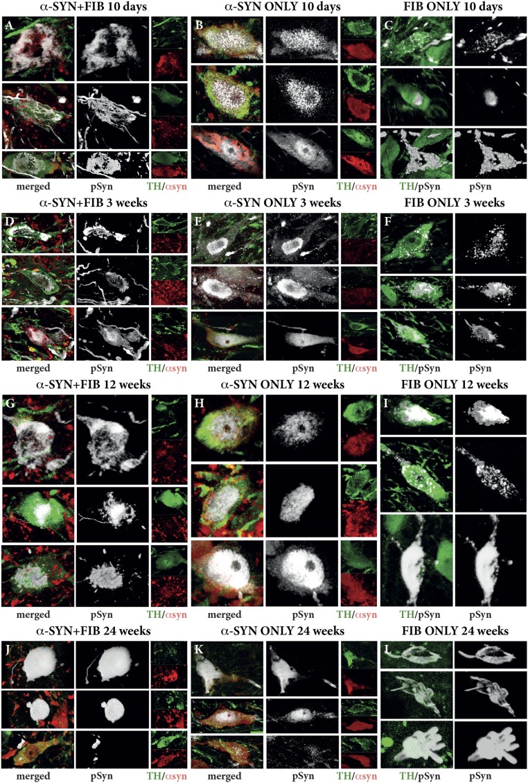
Fig. S3.
Development of Lewy-like synucleinopathy in nigral DA neurons over time. Triple-immunofluorescent staining shows the expression of the AAV-derived human α-syn protein (red) and the morphological appearance of the PFF-induced pSyn+ aggregates (white) in the TH+ neurons (green) in the SN. Representative photographs of individual neurons were selected from the α-SYN + FIB (Left), α-SYN ONLY (Center), and FIB ONLY (Right) groups at four different time points after PFF injection: 10 d (A–C), 3 wk (D–F), 12 wk (G–I), and 24 wk (J–L). Images were acquired as collated, 3D rendered images in the confocal microscope.

