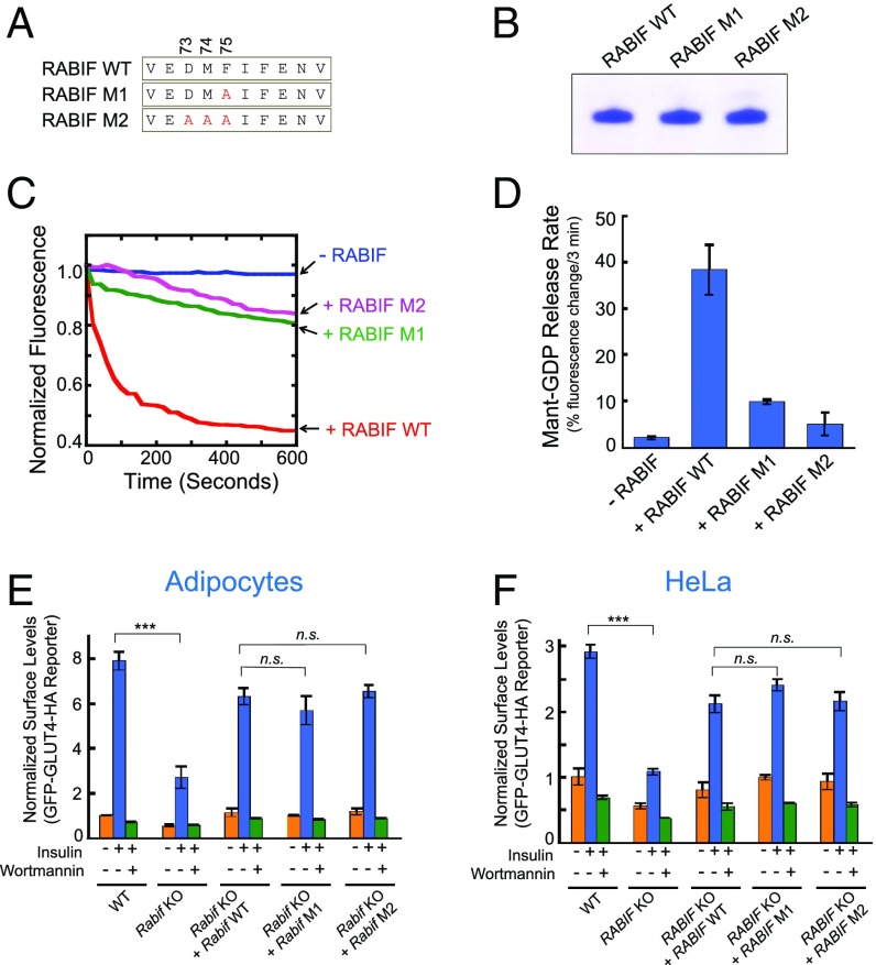
Fig. 4.
RABIF does not function as a GEF in GLUT4 exocytosis. (A) Diagram showing the RABIF point mutations predicted to impair its interaction with Rab10. (B) Coomassie blue-stained gel showing purified WT and mutant RABIF proteins. (C) Kinetics of fluorescence changes resulting from RABIF-catalyzed mant-GDP release. The reactions were carried out in the presence of WT or mutant RABIF, using prenylated Rab10 as the substrate. (D) Initial rates of the reactions in C. Data are shown as percentage of fluorescence change within the first 3 min of the reactions. Error bars indicate SD. (E) Normalized surface levels of the GLUT4 reporter in the indicated adipocytes. n.s., not significant. ***P < 0.001. Error bars indicate SD. (F) Normalized surface levels of the GLUT4 reporter in the indicated HeLa cells. ***P < 0.001. Error bars indicate SD.

