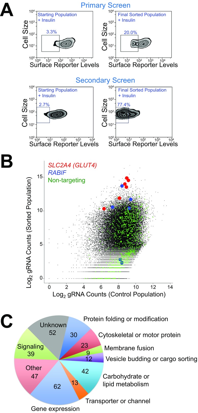
Fig. S2.
CRISPR-Cas9 genetic screens of insulin-stimulated GLUT4 exocytosis. (A) Flow cytometry analysis of the starting mutant population and the final sorted population in the genome-wide primary screen (Top) and the secondary screen (Bottom). HeLa cells were treated with 100 nM insulin for 30 min before the cells were stained and analyzed by flow cytometry. (B) Abundance of sgRNAs in the genome-wide screen and the passage control. (C) Summary of genes validated in the secondary screen.

