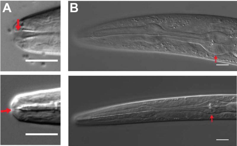
Figure 6. The non-feeding dauer stage is characterized by buccal cavity occlusion and pharyngeal bulb shrinkage.

DIC micrographs of L3 (top) and dauer (bottom) animals demonstrating changes to the buccal cavity (A, red arrow) and the pharynx (B). Shrinkage of the pharynx is most obvious in the terminal bulb (red arrow). Scale bars, 10 μm
