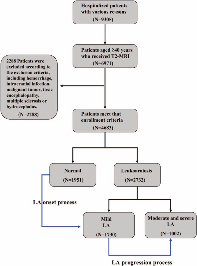
Figure 1.

Flowchart of subject recruitment for the study population in the First Hospital affiliated with Xiamen University, including residents 40 years and older who showed normal neuroimaging or white matter hyperintensity on the T2-weighted FLAIR MRI scans of brain scans. LA = Leukoaraiosis, MRI = magnetic resonance imaging.
