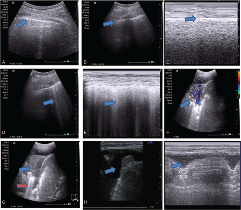
Figure 2.

Ultrasound findings. (A) Normal lung ultrasound showing the pleural line (blue arrow); (B) Normal lung ultrasound showing pleural ribs between artifacts (blue arrow); (C) M-type ultrasound normal pleural line; (D) sparse line B1 (blue arrow); (E) M-type fusion line B2 (blue arrow); (F) blood flow signals inside lung consolidation (blue arrow); (G) dynamic air bronchogram inside lung consolidation, in with lung consolidation (blue arrow) and sparse line B1 (red arrow) were observed; (H) pleural effusion (blue arrow); (I) M-type ultrasound line showing the pleural effusion lung line (blue arrow).
