Figure 1.

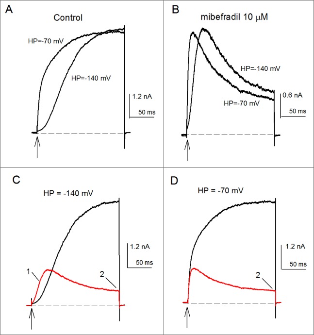
Mibefradil inhibition of the Cole-Moore shift and K+-conductance of Kv10.1 channels. (A) Control IK evoked by a 200-ms pulse to +30mV applied from the HP of either -70 or -140 mV, as indicated. The rate of IK activation depends on the HP from which channels are activated. IK activated from -140 mV is markedly slower than IK evoked from the HP of -70mV. (B) As in A, but in the presence of 10 µM mibefradil. (C) Superposed IK in control (black trace) vs. 10 µM mibefradil conditions (red trace). The currents were activated as in A from the HP of -140 mV. Notice the clear mibefradil inhibition of the Cole-Moore shift. As a consequence of this, the initial IK of mibefradil-modified channels surpasses control IK (labeled 1). Also note the marked mibefradil inhibition of GK at pulse end (labeled 2). (D) As in C but currents were activated from the relatively depolarized HP of -70 mV. Note that, as expected, in this case mibefradil only inhibits GK at pulse end. The vertical arrows indicate the time of delivery of the constant +30 mV activation pulse. The dashed line is the zero current level.
