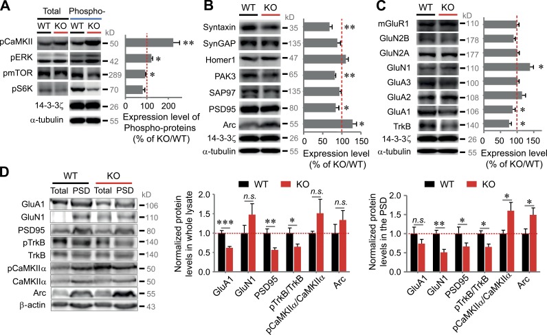
Figure 5.
Biochemical changes in the hippocampal PSD fraction and whole lysates of VRK3-KO mice. (A) CaMKIIα, ERK1/2 (p42/44), mTOR, and S6K proteins. The level of phosphoproteins was normalized by KO/WT ratios of total proteins (n = 5–7 for WT and VRK3-KO mice; **, P < 0.01; *, P < 0.05, t test). (B) Levels of syntaxin, SynGAP, Homer1, PAK3, Arc, SAP97, and PSD-95; all proteins were normalized by α-tubulin or/and 14-3-3ζ (n = 5–7 for WT and VRK3-KO mice; **, P < 0.01; *, P < 0.05, t test). (C) Levels of mGluR1, GluN1, GluN2A, GluN2B, GluA1, GluA2, GluA3, and TrkB; all proteins were normalized by α-tubulin or/and 14-3-3ζ (n = 5–7 for WT and VRK3-KO mice; *, P < 0.05, t test). The same 14-3-3ζ loading control was used in both B and C, being the same test group. (D) Immunoblot analyses of PSD fractions and whole lysates from 12–13-wk old WT and VRK3-KO mice for the indicated proteins (left). Protein levels of GluA1, GluN1, PSD-95, phosphorylated TrkB, phosphorylated CaMKIIα, and Arc in the hippocampal whole lysates (middle) and PSD fractions (right) from VRK3-KO mice (n = 6 mice per genotype; ***, P < 0.001; **, P < 0.01; *, P < 0.05, t test). β-Actin was used as a loading control, and all values were normalized to the mean level of the respective protein in the PSD fractions or whole lysates from WT mice. n.s., not significant. All values represent mean ± SEM.

