Figure 3.
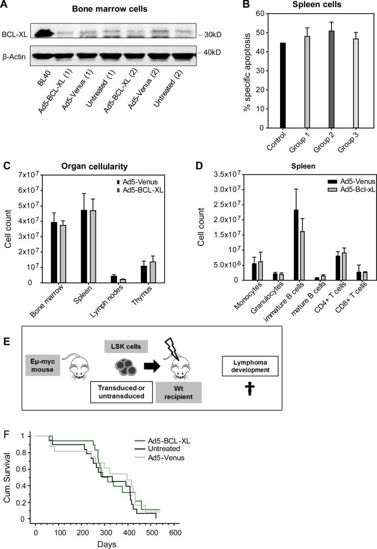
Adenoviral BCL-XL overexpression has no long-term adverse effects. (A) Recipient mice were transplanted as indicated in Fig. 2 A and sacrificed 16 wk later. Total BM cells were analyzed for their BCL-XL protein contents by Western blot. The Burkitt lymphoma cell line BL40 was used as positive control and β-actin as loading control. The legend indicates how the Ly5.2+ donor cells were treated before transplantation. (B) Splenic cells of recipient mice were cultured for 24 h in the presence or absence of etoposide (1 µg/ml), and specific apoptosis was determined by flow cytometry. Spleens of WT mice were used as a control. Bars represent means of n = 3 independent experiments ± SEM. No significant differences were observed (Mann–Whitney test). (C and D) Ly5.1+ recipient mice were transplanted with 15,000 Ly5.2+ LSK cells transduced with Ad5-Venus or Ad5–Bcl-xL. To ensure survival of the recipients, 150,000 Ly5.1+ BM cells were co-transplanted. Recipient mice were sacrificed 1 yr later and analyzed in detail. No signs of myelo- or lymphoproliferation or leukemia were observed in the indicated organs. Bars represent means of n = 6 animals ± SEM; no significant differences were observed (Mann–Whitney test). (E) LSK cells were isolated from the BM of Eμ-myc tg mice and left untreated or transduced with the Ad5-Venus or Ad5–BCL-XL adenoviruses. Subsequently, LSK cells were transduced into lethally irradiated recipient mice, which were monitored for lymphoma development. (F) When in poor general condition, recipient mice were sacrificed and analyzed. The Kaplan–Meier blot shows n = 13–18 mice per group (four independent experiments). No significant p-values between the groups were obtained by using the log-rank (Mantel–Cox) test.

