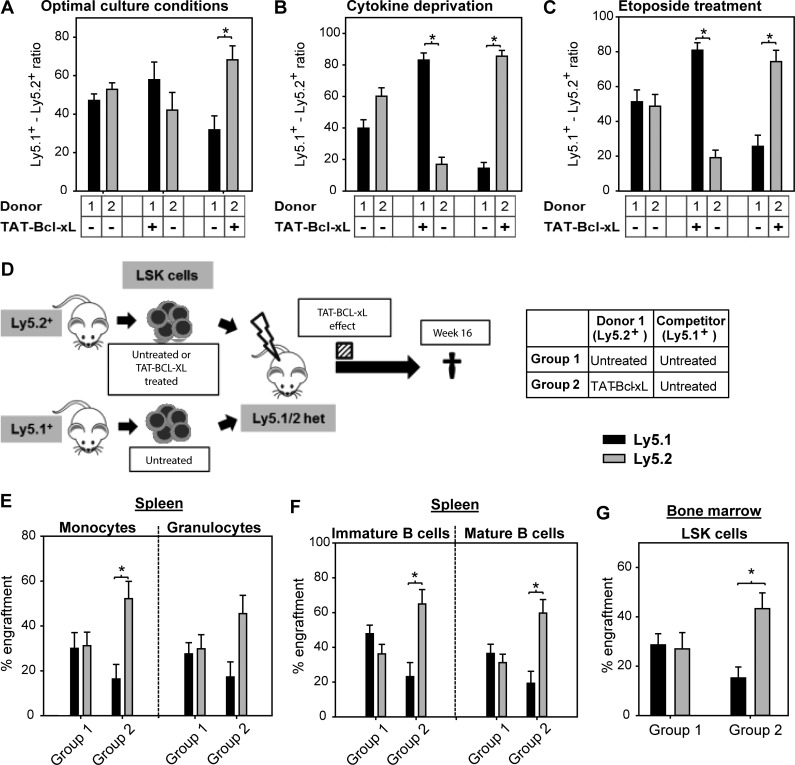
Figure 5.
TAT–Bcl-xL protein transduction increases competitiveness of LSK cells in vitro and in vivo. (A–C) Ly5.1+ and Ly5.2+ LSK cells were cultured in a 50:50 ratio under different culture conditions. Cells were either pretreated with TAT–Bcl-xL or left untreated and cultured for 5 d under optimal culture conditions (10% serum with SCF, TPO, and Flt3L, 100 ng/ml each; A), in the absence of cytokines (B), or in the presence of etoposide (C). Bars represent means of n = 4 independent experiments ± SEM. Significant p-values (Wilcoxon Test): P = 0.04 in A–C. (D) Ly5.2+ LSK cells treated either with TAT–Bcl-xL or left untreated were co-transplanted with untreated Ly5.1+ cells in a 50:50 ratio into lethally irradiated Ly5.1/Ly5.2 heterozygous mice. 16 wk later, recipient mice were sacrificed, and donor chimerism was determined by flow cytometry. The dashed bar indicates the period, in which Ly5.2+ LSK cells were containing TAT–Bcl-xL. (E–G) Chimerism was detected in splenic monocytes and B and T lymphocytes (E and F), as well as in BM-derived LSK cells (G). Bars represent means ± SEM of n = 10–11 mice from three independent experiments. Significant p-values (Wilcoxon test): (E) P = 0.02 for Group 2 in monocytes; (F) P = 0.03 for Group 2 in immature and mature B cells; (G) P = 0.03 for Group 2 in LSK cells.

