. 2017 Oct 3;6:e29738. doi: 10.7554/eLife.29738

Figure 2. Gadobutrol vs. gadofosveset in MRI-visualization of dural lymphatic vessels.

Coronal T1-weighted black-blood images were acquired after intravenous injection of two different gadolinium-based contrast agents (31 min after gadobutrol and 42 min after gadofosveset), during two MRI sessions separated by one week. Dural lymphatics (red arrows in magnified view boxes) were better discerned using gadobutrol (standard MRI contrast agent, which readily enters the dura) compared to gadofosveset (serum albumin-binding contrast agent, which remains largely intravascular) and were localized around dural sinuses, middle meningeal artery, and cribriform plate (white arrows). Notably, the choroid plexus (white arrows) enhanced less with gadofosveset than gadobutrol, whereas meningeal and parenchymal blood vessels (both veins and arteries) did not enhance with any contrast agent and appeared black. On conventional T1-weighted MPRAGE images, meningeal and parenchymal blood vessels enhanced with both contrast agents, more clearly with gadofosveset.

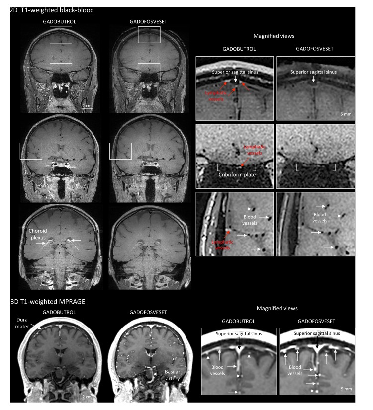
Figure 2.

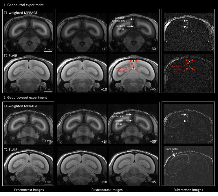
Figure 2—figure supplement 1. MRI visualization of dural lymphatic vessels in a healthy marmoset with different contrast agents.

Figure 2—figure supplement 1.

In an 11-year-old healthy common marmoset, high-resolution (150 × 150 μm in-plane) coronal T1-weighted MPRAGE and T2-FLAIR were acquired before and at different time points after the injection of two different contrast agents (single dose of gadobutrol and gadofosveset), a week apart. (1) Gadobutrol experiment. Dural lymphatic vessels are discriminated on postcontrast T2-FLAIR and on subtraction images (post vs. precontrast images), but not on postcontrast 3D T1-weighted MPRAGE. (2) Gadofosveset experiment. Gadofosveset, which binds serum albumin and therefore remains largely inside blood vessels (white arrows on postcontrast T1-MPRAGE and subtraction images), even in tissues (such as the dura mater) that lack a blood-brain barrier, is FDA-approved for magnetic resonance angiography. Dural lymphatic vessels are not visible on post-gadofosveset T2-FLAIR and relative subtraction images, even on delayed acquisition (45 min after injection), despite preserved enhancement of the dural venous sinuses. Numbers refer to minutes after intravenous administration of the contrast agent.