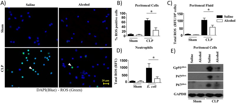
Fig 5. Attenuation of ROS production in alcohol-challenged mice and in alcohol-treated bone marrow-derived neutrophils in response to bacterial infection.
A, ROS+ neutrophils were identified by fluorescence microscopy after intracellular staining for ROS in peritoneal cells (neutrophils) derived from alcohol-administered and control mice at 24 h post-CLP. Results are representative of a microscopic view of three independent experiments (ROS+ cells are green, ROS− cells are blue). Arrows show ROS producing cells. B, Twenty random images were selected from three experiments and quantified for the presence of ROS-positive (green) neutrophils. (*, p<0.05). C, The production of total ROS in peritoneal fluid of alcohol-administered and control mice at 24 h post-CLP were evaluated as relative fluorescence units (RFUs) and normalized to total peritoneal cells. D. Total ROS production by bone marrow-derived neutrophils stimulated with E. coli were quantified as relative fluorescence units (RFU). (n = 5-8/group; *, p<0.05). E. Western blotting of GP91phox, P47phox, P67phox and GAPDH expression in peritoneal cells (neutrophils) from alcohol-administered and saline-challenged mice at 24 h post-CLP. This is a representative blot of 3 independent experiments with identical results. Original magnification 20x.

