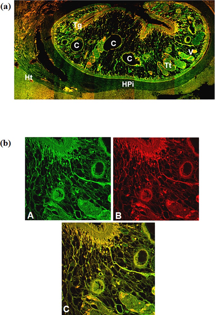
Fig 3.

Confocal micrograph of Fasciola gigantica in buffalo liver tissue section showing excretory secretory antigens in the parasite and the naturally infected host (Bubalus bubalis) liver as revealed by green fluorescence of FITC conjugated secondary antibody (a). The sections were counter stained with phalloidin TRITC (red fluorescence) for actin filaments. (b) Magnified micrograph showing immunolocalization of ES antigens using FITC (green) conjugated secondary antibody (A) and counter stained using phalloidin TRITC (B). The co-localization of antigens in the sub-tegumental and parenchymatous region as evident from the yellow fluorescence while green alone is showing exclusive antigen distribution (C). [C intestinal caecae, Tt testes, V vitellaria, Tg tegument, Ht host tissue and HPi host-parasite interface].
