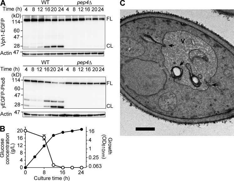
Figure 1.
Vacuolar dynamics after diauxic shift. (A) Immunoblot analyses of Vph1-EGFP and yEGFP-Pho8 expressed in either the WT or pep4Δ strain. Cells were taken from cultures in YEPD medium at 28°C for the indicated time period and subjected to immunoblot analysis with an anti-GFP antibody. FL represents a band position for the full-length form of the fusion proteins, and CL indicates that for the cleaved form containing EGFP or yEGFP moiety. The immunoblot data gained with anti–β-actin antibody are shown as loading controls. (B) Time course of glucose consumption (open circle) and growth (closed circle) of the WT strain in YEPD medium. The mean values from three independent cultures are plotted, and error bars indicate standard deviation. (C) A representative EM image of a WT strain cultured in YEPD medium at 28°C for 16 h. The vacuole invaginations are highlighted with black arrows. V, vacuole. Bar, 0.5 µm.

