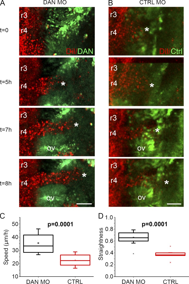
Figure 7.
In vivo neural crest cell speed, directionality, and proliferation are increased when DAN is knocked down in the paraxial mesoderm. (A) Selected images from a time lapse showing neural crest cells (red) migrating into the paraxial mesoderm transfected with DAN MOs (green). (B) Selected images from a time lapse showing neural crest cells migrating into paraxial mesoderm transfected with control MOs (green). Asterisks indicate the front of the neural crest migratory stream. Bars, 50 µm. (C) Speed of neural crest cells migrating into paraxial mesoderm transfected with DAN MOs (n = 13) and control MOs (n = 9). (D) Directionality of the same cells tracked in C. Two-sided Student’s t test. OV, otic vesicle.

