Figure 3.
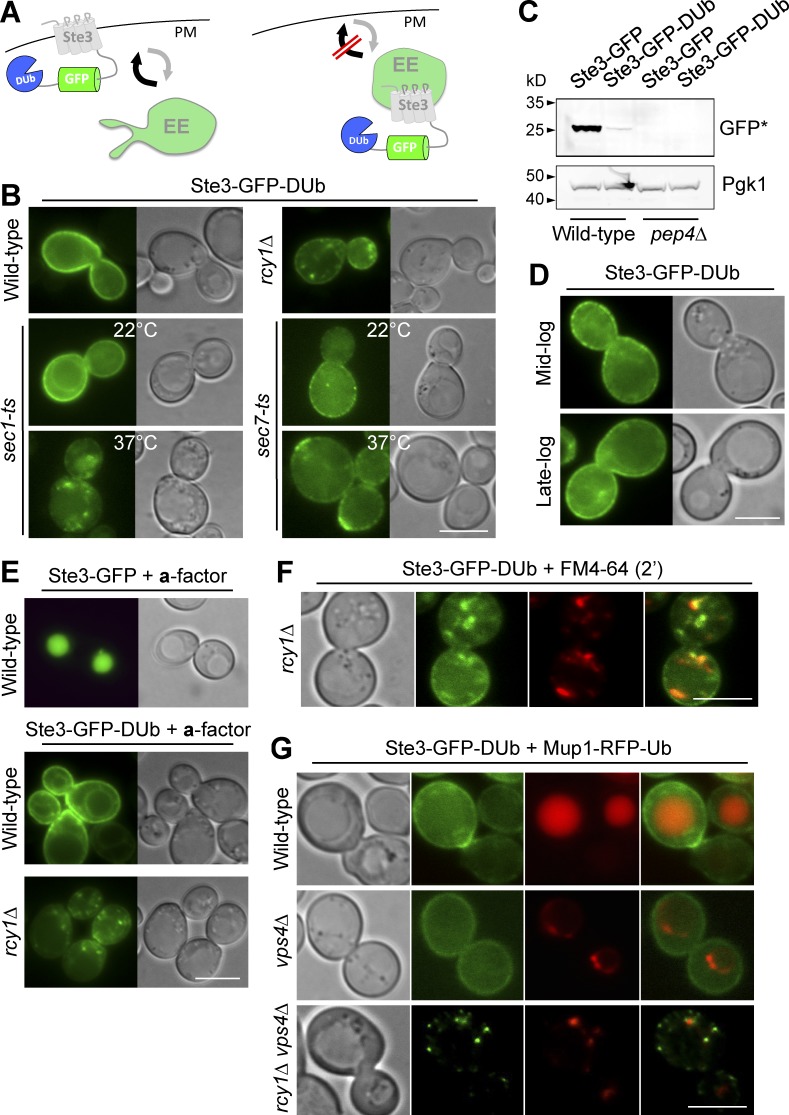
A synthetic reporter that follows an Rcy1-dependent EE>PM route. (A) Ste3-GFP-DUb, comprising the Ste3 G protein–coupled receptor fused to GFP and the catalytic domain of DUb, designed to be at the cell surface in wild-type cells and endosomes in recycling mutants. (B) Localization of Ste3-GFP-DUb in wild-type and rcy1Δ cells or sec1-1 (ts) and sec7-1 (ts) cells grown at 22°C and 37°C. (C) Immunoblot of vacuolar-processed GFP cleaved from Ste3-GFP-DUb or Ste3-GFP expressed in wild-type and pep4Δ cells. (D) Ste3-GFP-DUb localization at mid- and late-log phase. (E) Localization of Ste3-GFP and Ste3-GFP-DUb in labeled cells grown in medium enriched with a-factor. (F and G) Ste3-GFP-DUb expressed in indicated mutants colocalized with FM4-64 chased for 2 min (F) or Mup1-RFP-Ub in the presence of 20 µg/nl methionine (G). Bars, 5 µm.

