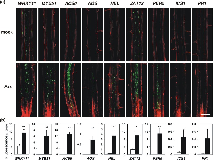
Fig 6. Expression of promoter::YFPN constructs in roots infected with F. oxysporum.
(a) Microscopic analysis of the infection site of 12-day old roots 2 days after inoculation with spores from F. oxysporum. Fluorescence derived from promoter::YFPN constructs is localized in the root nuclei and shown in green, while propidium iodide was used as a counterstain of cell walls and dead cells and is shown in red. Bar 100 μm. (b) Quantification of microscopic analysis of promoter::YFPN constructs as depicted in (a) using Fiji. Bars represent the mean of ≥ 6 images ± SE. Statistical analysis was performed using a Student’s t-test: * p < 0.05, ** p < 0.01, *** p < 0.001.

